


Kalpa Media House | Bengaluru(Whitefield) |
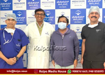
A remarkable case of misdiagnosed thyroid cyst that was later revealed to be a bronchogenic cyst has been successfully treated at Medicover Hospital, Bengaluru, underscoring the critical importance of accurate radiological diagnosis and the role it plays in guiding appropriate clinical management.
A 33-year-old homemaker, who had been struggling with chronic cough, dyspnea (shortness of breath) and general weakness for several weeks, initially sought medical attention at a local nursing home.
Also Read>> Sounds of melody at Madhuradhwani
Imaging results pointed to a large cyst (cyst is a benign growth containing liquid or semisolid material) located in the thyroid region, prompting further investigations. Fine-Needle Aspiration Cytology (FNAC) initially suggested a Colloid Cyst from Thyroid Gland with extension into chest (superior mediastinum), but after a sudden increase in the size of the mass, patient was referred to Medicover Hospital for more comprehensive evaluation and management.
At Medicover Hospital, further imaging, including CT and MRI scans, revealed that the mass measuring 8.2 x 7.5 x 5.4 cm did not arise from the thyroid but rather from the superior mediastinum. After a diligent re-evaluation by Consultant Radiologist Dr. Chaynika Agarwal, the cyst was diagnosed as a bronchogenic cyst – a rare congenital anomaly resulting from abnormal development of the tracheobronchial tree and was seen pushing the trachea to the left.
 This condition, though benign ( non-cancerous) is asymptomatic in many cases, but can cause symptoms like cough and dyspnea when it compresses adjacent structures such as trachea or bronchus.
The Cardiothoracic Team led by Senior Cardiothoracic Surgeon Dr. Raghavendrra Chikatoor opted for a surgical approach involving an upper hemisternotomy. The procedure, which required splitting the upper half of breast-bone and careful dissection due to the cyst’s attachment to the trachea, major blood vessels and nerves in the neck, was successfully performed without any complications. The cyst was removed in its entirety and histopathological examination confirmed the diagnosis of a non-pulmonary benign bronchogenic cyst.
Following the surgery, the patient experienced immediate relief from her symptoms. Two weeks after her discharge, she reported a significant improvement in her quality of life, with complete resolution of cough and dyspnea that had plagued her for weeks.
 Dr. Raghavendrra Chikatoor emphasized the significance of this case, stating, “This case serves as a reminder of the critical role radiological imaging plays in accurate diagnosis. Had the diagnosis remained a thyroid cyst, this patient might have undergone unnecessary thyroid surgery, which would have impacted her thyroid function for life. Instead, a clear understanding of the cyst’s true origin allowed us to preserve her thyroid gland and its function while addressing the compression on her trachea. We also could avoid an additional incision in the neck.”
Dr. Chaynika Agarwal added, “This case highlights the importance of a multidisciplinary approach, as re-evaluation and collaboration between radiology and surgery ultimately led to the correct diagnosis and an optimal surgical outcome.”
This case exemplifies the life-changing potential of accurate diagnostics, careful surgical intervention and a collaborative medical approach.
Contact for news and advertising: Whatsapp: 9008761663, 9481252093 – info@kalpa.news


